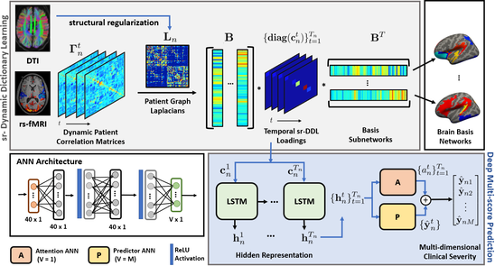
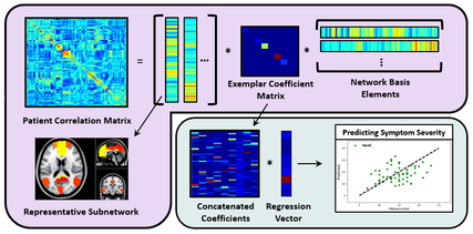
Resting State fMRI is a non-invasive neuroimaging modality which has become ubiquitous for studying neurological disorders such as Autism, ADHD and Schizophrenia. The individual impairment in social and cognitive functioning among clinical populations is typically quantified by biomarkers of symptom severity. These measures are assessed and scored by a trained clinician. Owing to the well known group level confounds of rs-fMRI, a reliable characterization of behavioral heterogeneity from neuroimaging data at an individual level is extremely challenging.
In this project, we develop a novel optimization framework which jointly models the group level and patient specific resting state information through a joint matrix factorization across patients. Going one step further, we use the patient level description to predict clinical severity via a regression model. We combine the neuroimaging representation and severity prediction steps into a single Joint Network Optimization (JNO) framework, and leverage computationally efficient optimization techniques to infer the latent representations inherent to our model. This helps us isolate key resting state co-activation signatures associated with various neuropsychiatric disorders, and, at the same time, map the wide spectrum of clinical manifestations across patients. Project Members : Dr. Mary Beth Nebel, Dr. Stewart Mostofsky, Dr. Archana Venkataraman [code] [paper] |
Improving Rehabilitation in Spinal Cord Injury Patients
Spinal cord injury (SCI) is a devastating trauma that can result in permanent loss of movement and sensation. While medical advancements have reduced the mortality rates for these patients, the quality of life for SCI patients remains relatively grim, as more than 50% of them will be left with severe paralysis, and the remaining will only partially recover their original functionality. Physical therapy and rehabilitation expedite recovery by retraining neural processes in the brain, a phenomenon known as functional reorganization. However, functional reorganization is not well characterized or understood, which limits both the development of more targeted therapy plans and the extraction of biomarkers for recovery.
Resting-state fMRI has the potential to detect functional reorganization due to SCI, which is necessary to identify therapeutic targets and track treatment induced changes. Combining machine learning theory with neuroimaging data and clinical insights can help explain functional reorganization of patients who sustained a spine or brain injury. We have begun to identify whole-brain connectome in these patients using prior established methods from our work. Namely, we are pinpointing specific degraded functional connections using a matrix factorization framework and characterizing subject-specific functional organization using a Bayesian approach. Project Members: Naresh Nandakumar, Dr. Ann Choe, Dr. Archana Venkataraman Self -Taught Learning based Optical Fluorescence Microscopy Image Deblurring using Stacked Denoising Auto-encoders
Fluorescence microscopy is an essential part of a biologist’s toolkit, allowing assaying of many parameters like sub-cellular localization of proteins, changes in cytoskeletal dynamics, protein-protein interactions, and the concentration of specific cellular ions. A fundamental challenge with studying tissue structures at a specific depth using fluorescence microscopy is the presence of noise from layers at other depth and the lack of denoised ground truth images.
We developed a framework to tackle the problem of deconvolution in Optical Fluorescence Microscopy Images using Self Taught Learning. This technique entailed developing and training a stacked auto-encoder architecture on Image-Net data set followed by a domain adaptation onto bright field microscopy images having ground truth available. Finally, the learned representations we deployed onto the optical fluorescence microscopy domain for deblurring in a transfer learning setting. Project Members: Rachana Sathish, Abhijit Guha Roy, Dr. Debdoot Sheet |
